🔬 Color Deconvolution: Unmix Biological Stains with Precision
PhenoCapture AI’s Color Deconvolution tool is a highly specialized, game-changing feature for digital pathology and biological image analysis. It mathematically separates brightfield histology images into individual stain channels (such as Hematoxylin, Eosin, or DAB) based on their specific optical density (OD) absorbance.
Whether you are performing quantitative tissue analysis, evaluating protein expression in IHC slides, or simply trying to isolate cell nuclei from a busy background, this tool instantly unmixes complex overlapping colors using the industry-standard Ruifrok & Johnston algorithm.
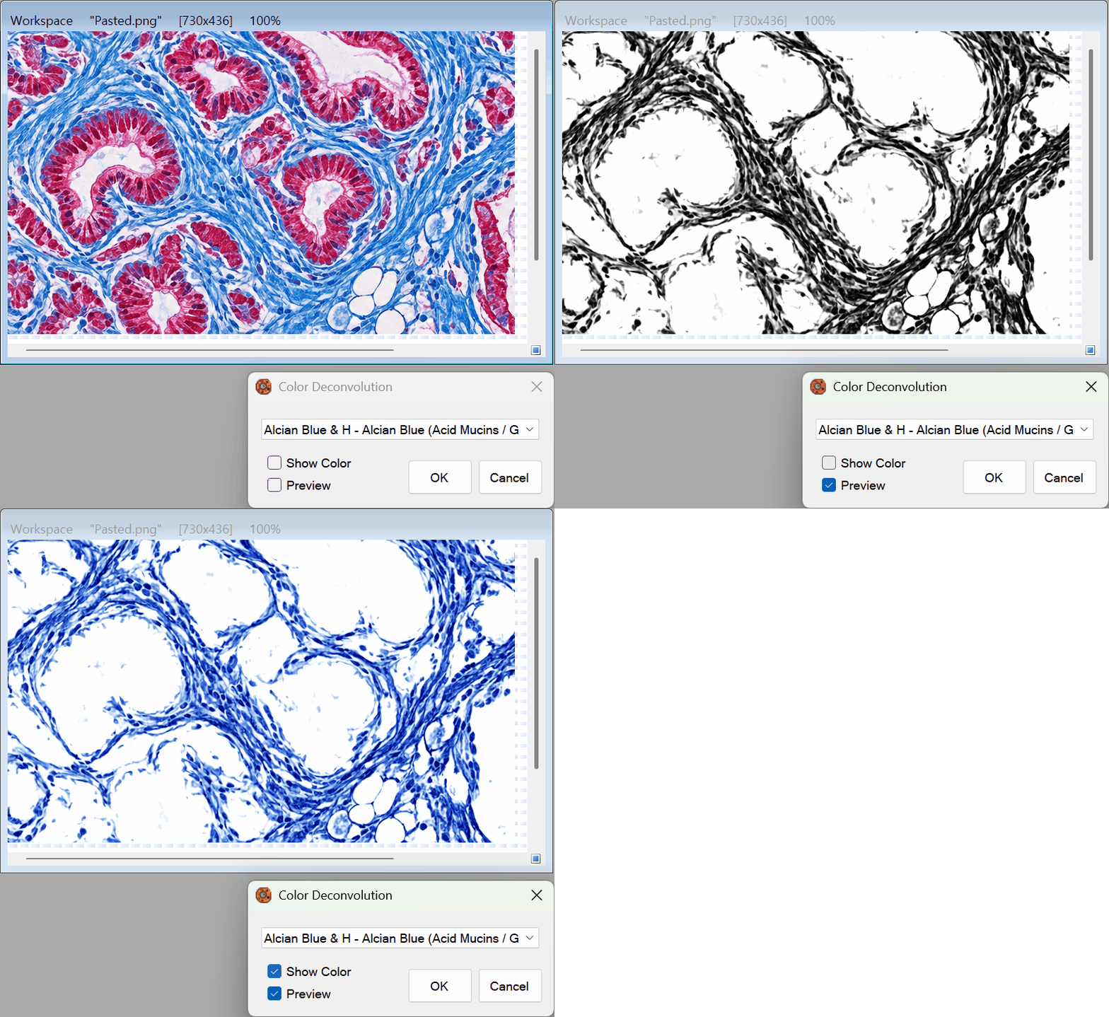
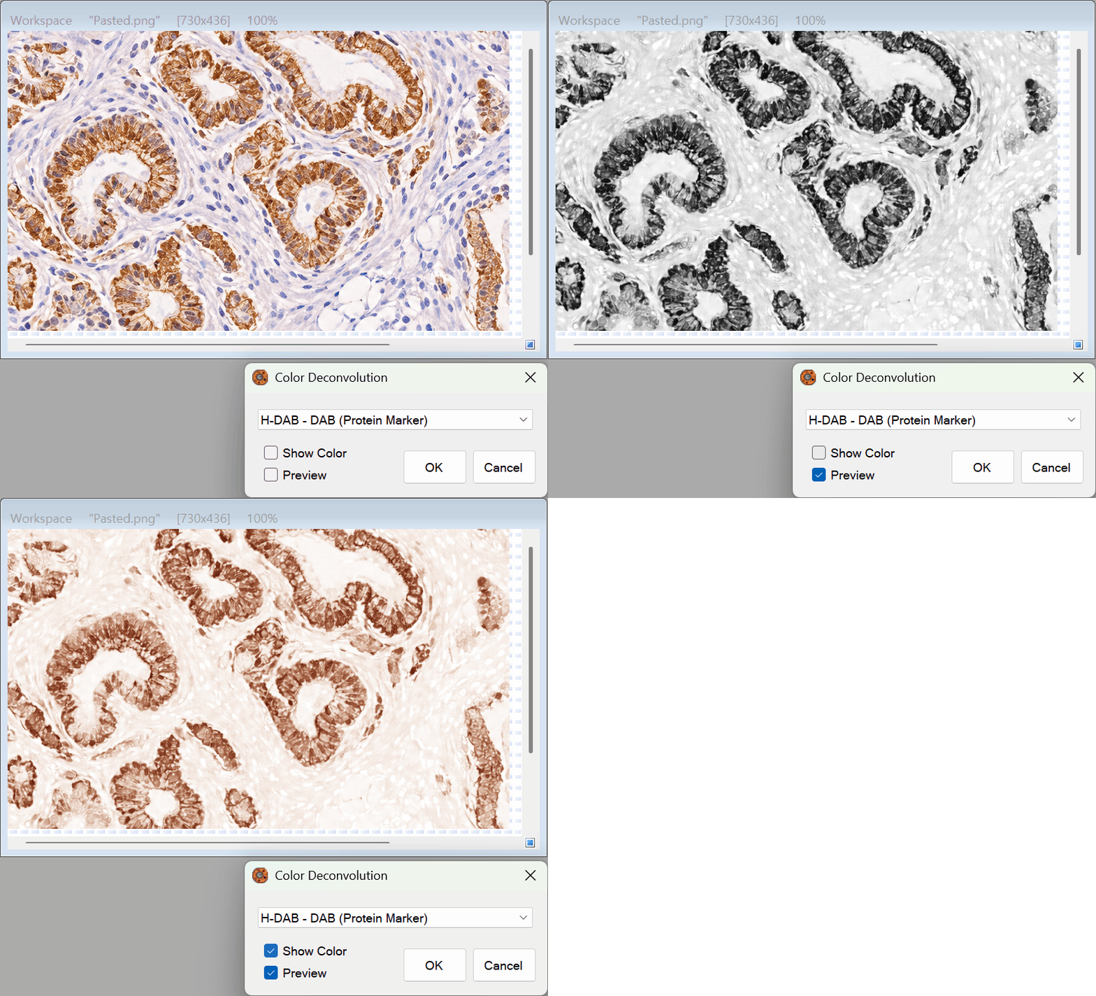
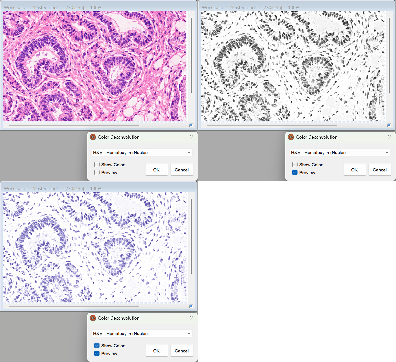
💡 Key Features
- 23 Built-In Stain Presets: Instantly deconvolve all major biological and histological stains. The software includes highly accurate OD matrices for H&E, H-DAB, Masson’s Trichrome, PAS, Giemsa, Alcian Blue, Feulgen, and even complex triple stains like H&E-DAB.
- Pseudo-Coloring (Show Original Color): Traditional deconvolution outputs a flat grayscale concentration map. With our advanced Show Original Color toggle, the engine performs a color re-convolution, allowing you to visualize the isolated stain exactly in its natural, brilliant color (e.g., viewing only the pink Eosin cytoplasm while hiding the blue nuclei).
- Real-Time Preview Engine: Instantly see the results of your stain separation on the canvas before committing. Switch between different target markers seamlessly with zero lag.
- Multi-Core Parallel Processing: Engineered with a highly optimized Optical Density (OD) Look-Up Table (LUT) and parallel thread processing. Even massive, high-resolution microscope slide images are deconvolved in fractions of a second across all available CPU cores.
- Targeted ROI Masking: Need to quantify a stain only within a specific tumor region? Use our selection tools to draw a boundary. The algorithm will isolate the stain exclusively within your selected mask, leaving the rest of the image completely untouched.
🛠️ How to Use the Tool
Extracting pure stain channels from your histological images is fast and intuitive:
Step 1: Select Your Stain Type
Open the Color Deconvolution tool and choose your specific staining method from the dropdown menu (e.g., H&E – Hematoxylin (Nuclei) or H-DAB – DAB (Protein Marker)). Ensure the Preview checkbox is enabled to watch your image transform immediately.
Step 2: Choose Your Display Mode
Use the Show Original Color checkbox to define how you want to view the separated stain:
- Checked: Displays the isolated tissue structure in its natural histological color (ideal for visual presentation and intuitive analysis).
- Unchecked: Displays a pure mathematical concentration map in grayscale (ideal for strict thresholding, morphological measurements, and intensity quantification).
Step 3: Apply the Deconvolution
Once you are perfectly satisfied with the isolated stain in the preview, simply click the OK button. The overlapping background colors will be permanently removed, leaving you with a perfectly separated channel ready for deep scientific analysis!
Empower your pathology research with PhenoCapture AI’s Color Deconvolution—bringing invisible cellular structures into sharp, quantifiable focus!